
Figure 2.
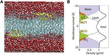
Melittin partitioned into the polar headgroup region of the lipid bilayer. (A) Snapshot of the simulation cell showing two melittin molecules (MLT1 and MLT2, in yellow) at the lipid-water interface. (B) Density cross-section of the simulation cell extracted from the 17-μs simulation. The peptides are typically located below the lipid phosphate (PO4) groups, in a similar depth as the glycerol/carbonyl (G/C) groups.