Fig. 5.

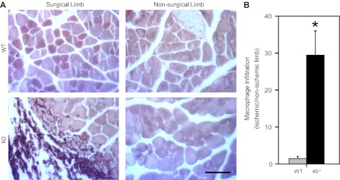
Macrophage infiltration was extensive in the Cx40−/− ischemic hindlimb. A: representative images of gastrocnemius muscle (in cross-section) isolated from mice 14 days after severe surgery and stained to reveal F4/80 antigen, a hallmark of activated macrophages (scale bar = 100 μm). Activated macrophages, stained dark brown, were more obvious in the surgical limbs of Cx40−/− mice. B: macrophage infiltration, quantified as the area of the surgical vs. nonsurgical gastrocnemius occupied by activated macrophages, was significantly (*P < 0.01) greater in Cx40−/− (n = 6, 4 male) compared with WT (n = 4 male) mice. KO, knockout mice.
