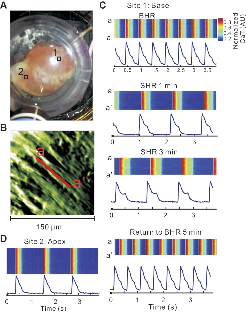
Fig. 5.
SHR promotes subcellular SCRs and CaT prolongation at the base but not the apex. A: locations of sites 1 and 2 (RVB and LVA, respectively) is indicated on this photograph of anterior wall of the heart. The high-magnification tracings from these sites are displayed in the other panels. B: subcellular image taken with the ×40 objective from site 1 at the RVB, as indicated in A. C: Cai transients recorded along line a–a′ in RVB shown in B at different HR in the line scan format along with tracings from the pixel located in the middle of line a–a′. Note the increasingly more pronounced SCR in SHR. D: Cai transients recorded along the longitudinal axis of a fiber in LVA at SHR.

