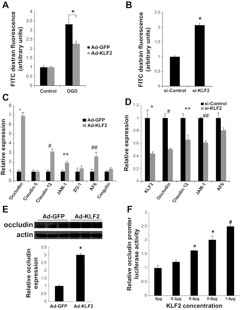
Fig. 6.
KLF2 regulates BBB permeability and occludin expression in brain microvascular ECs. A: transwell assays quantifying permeability of FITC dextran across primary human brain microvascular ECs infected with KLF2 (Ad-KLF2) or control (Ad-GFP) adenovirus at baseline and after 30 min oxygen glucose deprivation (OGD) followed by 2 h of normal media (n = 8/group). *P = 0.005. B: Transwell assays as in A using cells transfected with small interfering (si)-RNA (si-Control or si-KLF2) assessed at baseline (n = 8/group). *P < 0.0005 vs. si-Control. C: quantitative RT-PCR analysis of tight junction factors in human brain microvascular ECs infected with control (Ad-GFP) and KLF2 (Ad-KLF2) adenovirus (n = 3/group). *P < 0.0001, #P < 0.005, **P < 0.01, ##P < 0.05. D: quantitative RT-PCR analysis of tight junction factors in human brain microvascular ECs infected with si-RNA (si-Control or siKLF2) (n = 3 to 4/group). *P < 0.0001, #P = 0.01, **P = 0.01, ##P < 0.0001. E: Western blot analysis of occludin in human brain microvascular ECs infected with Ad-GFP or Ad-KLF2. Quantification of Western blot is shown. *P < 0.0001. F: transient transfection of 293T cells with occludin promoter luciferase reporter and increasing concentrations of KLF2 plasmid (n = 3/group). *P < 0.005, #P < 0.0005 vs. 0 μg. Representative results of 3 independent experiments shown.

