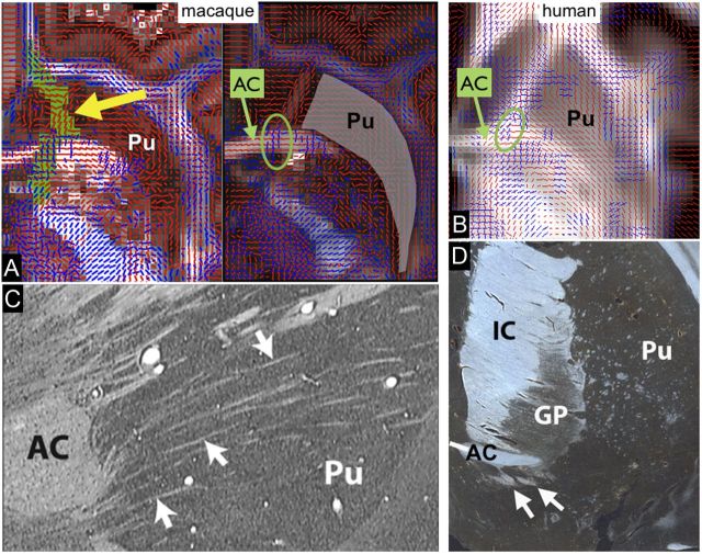
Figure 9.
A, Fiber orientations from dMRI in the vPFC. Two axial slices of the macaque brain (1 mm apart) showing fiber orientations estimated from BedpostX (red represents main orientation; blue, secondary orientation). The trajectory of the vmPFC connections from the chemical tracers data passes through a narrow passage within the striatum with noisy fiber orientations (tract shown in green and indicated by a yellow arrow). There are crossing fibers at the level of the AC at the exact location of the tracer's route. This result is in strong support of the model selection in BedpostX between one and two fibers per voxels. B, Axial slice of the human brain displaying fiber orientations from BedpostX. Similar to the macaque, crossing fibers are evident in the AC (green circle) at the level of the IC intersection. C, Nissl-stained sagittal section through the macaque brain. Arrows indicate small WM fascicles passing through the ventral striatum. D, GAP-43-stained coronal section through a human striatum. Arrows indicate WM fascicles passing through the ventral striatum. GP, Globus pallidus; Pu, putamen.

