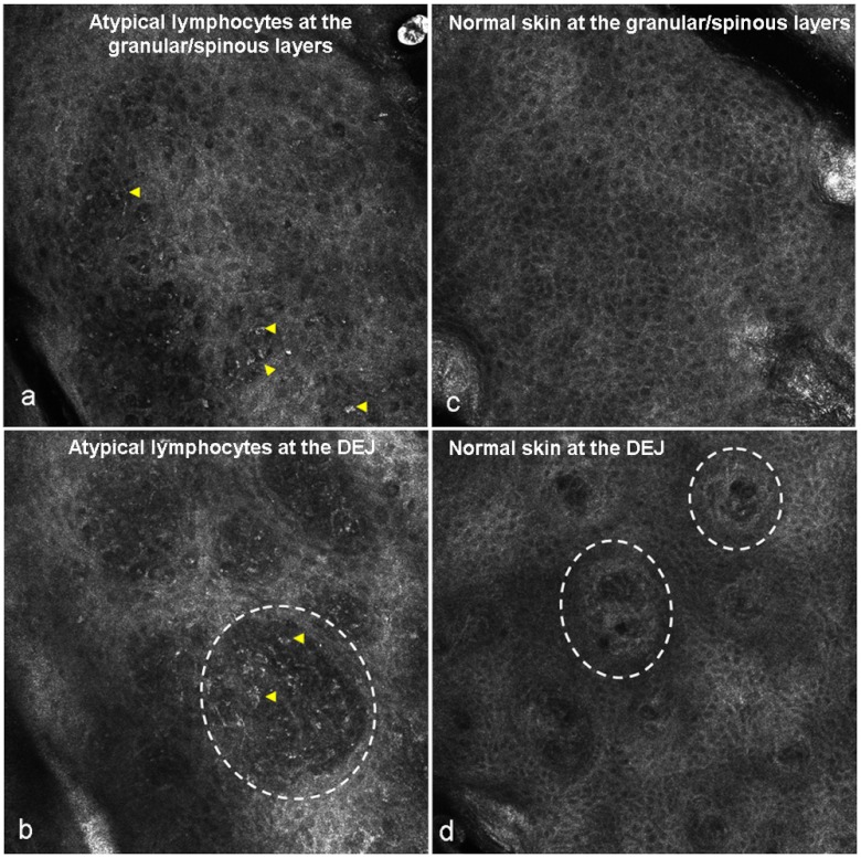
Fig. 1.
Panels (a) and (b) illustrate the presence of atypical lymphocytes in MF. Panel (a) demonstrates the presence of small to medium-sized bright round cells (yellow arrowheads) in between keratinocytes, which corresponds to exocytosis of lymphocytes on histopathologic examination. Panel (b) shows multiple bright round cells of varying morphology (yellow arrowheads) at the (DEJ within the dermal papillae (white dashed circle). Panel (c) illustrates normal honeycomb pattern of the granular/spinous layer, and (d) shows the DEJ morphology with dermal papillae (white dashed circle) in absence of any inflammatory infiltrate.

