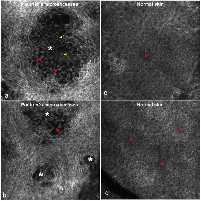
Fig. 5.
Panels (a) and (b) show representative images of Pautrier’s MF in mycosis fungoides, illustrating multiple, well-circumscribed, vesicle-like areas of varying size and dark reflectance (asterisk) at the granular-spinous layers. Within these nonrefractile areas, remaining keratinocytes can be visualized (red arrowheads). These keratinocytes focally still show adherence by intercellular connections, but intercellular spaces are widely enlarged and the epidermal structure is disrupted. Furthermore, small bright cells representing inflammatory cells are noted (yellow arrowheads). Panels (c) and (d) demonstrate corresponding RCM findings of normal skin at the granular-spinous layers with typical honeycomb pattern. Areas that appear slightly darker than the surrounding epidermis represent areas with underlying dermal papillae.

