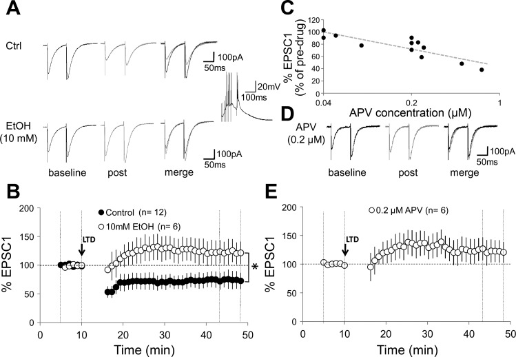
Fig. 6.
Parallel fiber long-term depression (LTD) [paired parallel fiber (PF) and CF activation] is blocked by ethanol in adult mouse Purkinje cells. A: typical traces in 0 mM Mg2+. Top left: PF-EPSCs before tetanization under control conditions. Top middle: PF-EPSCs 30 min after tetanization. Top right: overlay. Bottom traces: corresponding PF-EPSCs in the presence of 10 mM EtOH. Inset: tetanization pattern (8 PF pulses at 100 Hz, followed by single-pulse CF stimulation). This pattern was applied at 1 Hz for 5 min. B: time graph showing LTD under control conditions (n = 12) and LTD blockade in the presence of 10 mM EtOH (n = 6). C: d-APV blockade of CF-EPSCs is concentration dependent. CF-EPSCs were recorded in the presence of NBQX (10 μM). Graph plots the %change in EPSC amplitude against the concentration of d-APV (range: 0.04 to 1 μM). D: typical traces showing PF-EPSCs in the presence of 0.2 μM d-APV before (left) and 30 min after tetanization (middle). Right: overlay of traces. E: time graph showing an impairment of LTD in the presence of 0.2 μM d-APV (n = 6). Arrow indicates the time point of tetanization. Error bars are means ± SE. *P < 0.05, significant differences between the control and EtOH group by Mann-Whitney U-test.

