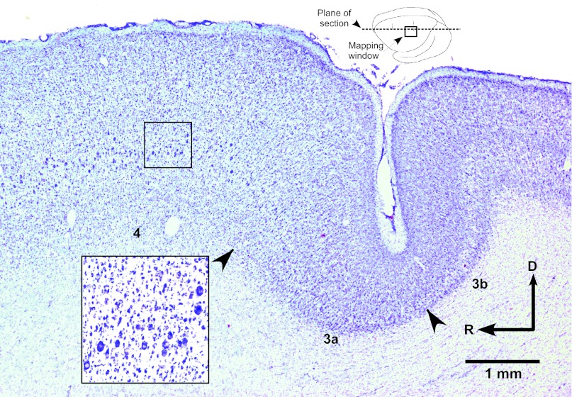
Fig. 3.
Photomicrograph of cresyl violet-stained parasagittal section through the M1 DFL representation. Inset at top shows plane of section in dorsolateral view of brain. Movements were evoked at ≤30 μA at ∼1,750 μm from the cortical surface within cortical regions containing large pyramidal cells in layer V, indicative of M1 (see inset at higher magnification). Cytoarchitectonically defined boundaries are indicated by arrowheads. D, dorsal.

