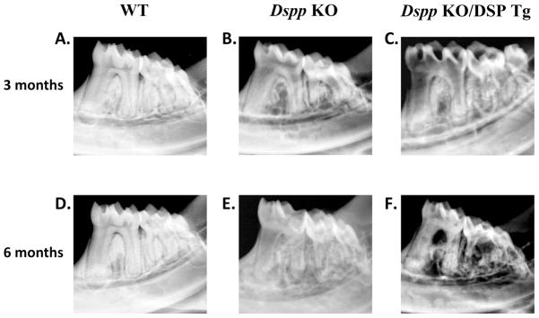
Figure 4. Flat X-ray radiography analyses of mandibles from 3- and 6-month-old mice.

At 3 and 6 months of age, the WT mice (A and D) showed evenly distributed and well mineralized radio-opaque dentin with small pulp chamber. The mandibular molars in the Dspp KO mice (B and E) had an enlarged pulp chamber and thinner dentin compared to the WT mice. The tooth defects in the Dspp KO/DSP Tg mice (C and F) were worse than those of the Dspp KO mice, with much thinner, radiolucent dentin and a more significant loss of the alveolar bone.
