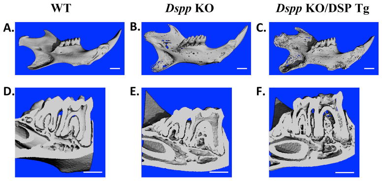
Figure 5. μ-CT analyses of mandibles from 3-month-old mice.

WT (A and D), Dspp KO (B and E) and Dspp KO/DSP Tg mice (C and F) were evaluated for their overall morphology (A, B and C) and dentin phenotypes in the molar cross sections (D, E and F). The WT mice showed evenly distributed and well mineralized dentin (A and D). The mandible of the Dspp KO mice (B) had porosities when compared to the WT sample. Molar cross section of the Dspp KO mice (E) showed an enlarged pulp chamber and thinner dentin compared with the WT mice (D). The defects in the Dspp KO/DSP Tg mice (C and F) were even more pronounced with marked increase in porosities and reduction in dentin thickness compared to the Dspp KO mice. Bar: 1 mm
