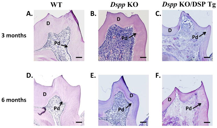
Figure 6. H&E staining in 3- and 6-month-old mice.
At 3 and 6 months, the Dspp KO mice (B and E) had wider predentin (denoted as “Pd”) and thinner dentin (denoted as “D”) compared to the WT mice (A and D). The Dspp KO/DSP Tg mice (C and F) displayed more severe dentin abnormalities compared to those of the Dspp KO mice (B and E). The dentin of the Dspp KO/DSP Tg (C and F) mice was almost non-existent with almost the entire layer consisting of the unmineralized predentin structure. The pulp showed evidence of ectopic calcification (C and F), which appeared worsened at 6-months (F) than 3 months of age. This could possibly be due to chronic inflammation arising from pulp exposure. All the six sections showed in this figure were processed, sectioned, stained and visualized using the same settings and protocols. The differences in the staining intensity seen in the sections were probably due to the difference in the matrix composition (e.g., increased amounts of proteoglycans in the Dspp KO/DSP Tg and Dspp KO mice). Bar: 100 μm.

