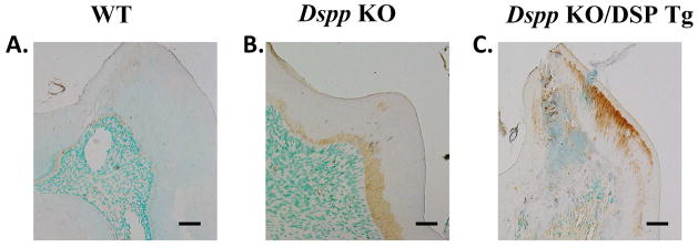
Figure 8. Biglycan immunostaining of predentin in 3-month-old mice.

The WT mice (A) showed a narrow predentin zone demarcated by the brown positive biglycan staining. In the Dspp KO mice (B), anti-biglycan activity revealed a widened predentin layer and an irregular dentin-predentin border as compared to the WT mice (A). The results from the Dspp KO/DSP Tg mice (C) indicated that nearly the entire predentin-dentin complex in these mice was made of the unmineralized predentin with anti-biglycan staining extending and covering most of the structure. Bar: A-H = 100 μm.
