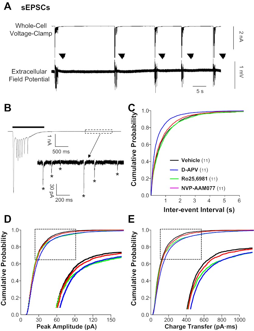
Fig. 1.
Spontaneous excitatory postsynaptic currents (sEPSCs) in granule cells were increased in hippocampal slice cultures treated chronically with N-methyl-d-aspartate receptor (NMDAR) antagonists. Whole cell voltage-clamp recordings were conducted at a −70-mV holding potential in physiological recording buffer containing bicuculline methiodide (BMI; 10 μM) and pipette solution containing QX-314 (5 mM) to block action potentials. Cumulative probability plots were generated from events pooled from all cells. A: simultaneous whole cell voltage-clamp (top) and extracellular field potential recordings (bottom) show BMI-induced epileptiform burst activity (arrowheads) in a dentate granule cell and dentate gyrus, respectively, in a vehicle-treated hippocampal slice culture. B: expanded time scale shows a whole cell voltage-clamp recording of a large-amplitude, long-duration sEPSC (sEPSClarge; line) associated with an epileptiform burst. The boxed area after the burst is expanded in the inset showing individual small-amplitude sEPSCs (sEPSCsmall; *). Cumulative probability plots show increased sEPSC frequency (C) in granule cells from slice cultures treated with d-aminophosphonovaleric acid (d-APV) compared with all other treatment groups and increased sEPSC amplitude (D) and charge transfer (E) for d-APV and Ro25-6981 compared with vehicle. Insets in D and E are expansions of regions enclosed in boxes. Legend in C applies to C–E; the number of granule cells/slice cultures is indicated in parentheses. See Table 1 for means.

