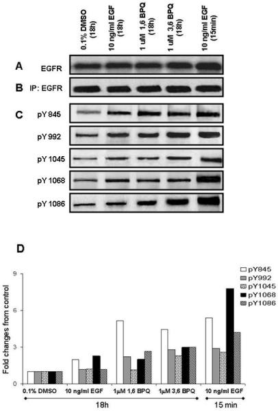
Figure 2.
Effect of BPQs on phosphorylation of the EGFR phosphosites under growth factor-dependent conditions. (A) Cell lysates were prepared in lysis buffer, subjected to Western Blot analysis. (B, C) EGFR was immunoprecipitated from MCF-10A cells treated as was specified in “Material and Methods”. Immunocomplexes were separated by SDS-PAGE and subjected to Western blot analysis for EFGR phosphorylation using an anti-EGFR or site-specific antibodies as indicated. (D) Densitometric analysis of the bands was used in order to make a semi-quantitative analysis of phosphorylation of EGFR phosphosites. The relative phosphorylation levels were estimated as compared to control. These results are representative of several independent replicates performed for each of the different phosphorylation sites.

