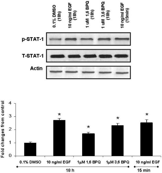
Figure 4.
STAT 1 activation is induced by BPQs in MCF-10A cells. Representative blot and quantified data show that STAT-1 phosphorylation in MCF-10A cells in media without EGF treated with 1 μM BPQs or 10 ng/ml EGF for 18 hr. Cell lysates were blotted with phosphotyrosine antibody to determine the phosphorylation status of STAT-1, the membrane was stripped of signal, and total STAT-1 levels were determined. 10 ng/ml EGF (15 min exposure) is included as positive control. Statistical analyses are for this individual experiment. Values are means ± S.E. * p< 0.05 as compared with control group.

