Figure 2.

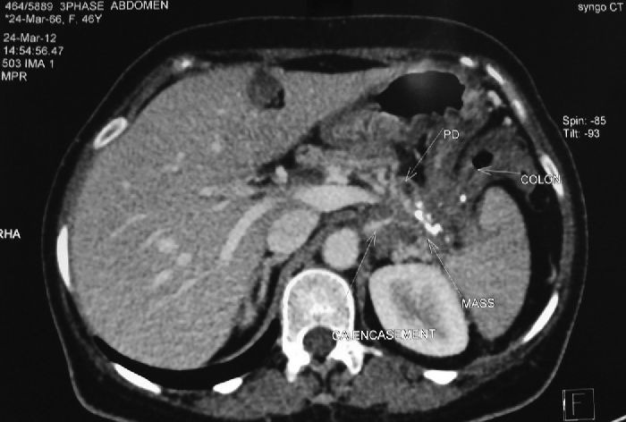
Computerized tomography abdomen of patient-2 showing atrophic pancreas, multiple calculi in the tail of pancreas along with a 46 mm × 34 mm × 31 mm encasing the celiac plexus infiltrating the splenic hilum and splenic flexure of colon

Computerized tomography abdomen of patient-2 showing atrophic pancreas, multiple calculi in the tail of pancreas along with a 46 mm × 34 mm × 31 mm encasing the celiac plexus infiltrating the splenic hilum and splenic flexure of colon