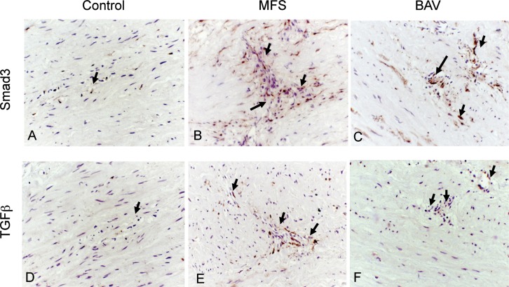
Figure 1.
Increased accumulation of Smad3 (A-C) and transforming growth factor beta (TGF-β) (D-F) observed in the area of pathological remodeling/focal degeneration in the aortic wall of Marfan syndrome (MFS) (B, E) and bicuspid aortic valve (BAV) (C, F) aneurysms. Positive expression of Smad3 and TGF-β is identified as areas of dark brown color (arrows). The control aortic wall (A, D) showed weak to negligible expression (hematoxylin;magnification 250×).

