
Figure 5.
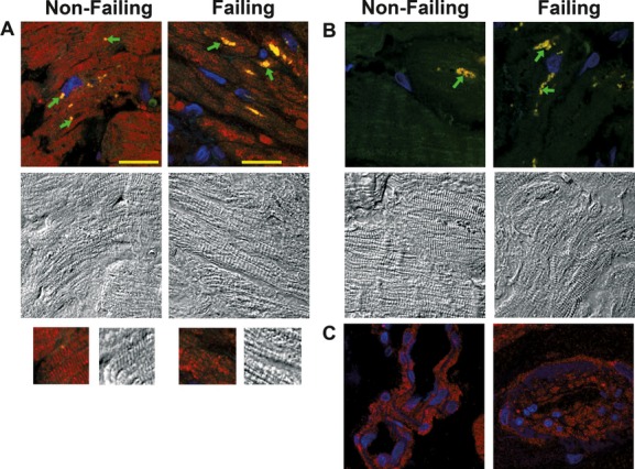
Anti‐KCNN2 immune reactivity in human cardiac tissue. A, Color and black‐and‐white panels show confocal fluorescence images and differential interference contrast (DIC) images, respectively, taken from nonfailing and failing hearts stained for KCNN2. Arrows denote areas of yellow fluorescence that were also detectable in the absence of the secondary antibody and result from the overlap of unspecific red and green autofluorescence in the tissue from nonfailing and failing hearts. B, Confocal fluorescence and DIC images taken from sections that were incubated with the secondary antibody only. C, Anti‐KCNN2 immunofluorescence in small‐artery walls. Scale bar, 20 μm.