
Figure 3.
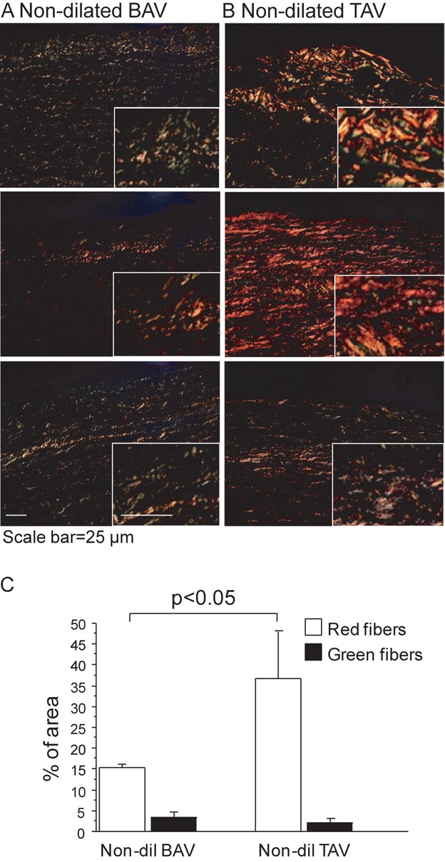
Picrosirius red staining of nondilated aortas from 3 randomly selected patients with BAV (A) and TAV (B). Green fibers indicate immature thin fibers and orange‐red fibers indicate mature, thicker, and better‐aligned collagen fibers. Magnifications of the stained areas are shown in right corner in each figure. All figures have been taken using the same conditions and settings. Scale bar=25 μm. C, Quantification of picrosirius red staining. Error bars are indicated by SEM. BAV nondilated (Non‐dil), n=3; TAV nondilated (Dil), n=3. BAV indicates bicuspid aortic valve; TAV, tricuspid aortic valve.