
Figure 4.
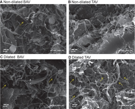
Scanning electron microscopy of collagen structure in ascending aortas. Nondilated aortas from patients with BAV (A) and TAV (B); dilated aortas of patients with BAV (C) and TAV (D). Representative photomicrographs from a total of 10 patients are shown. Thin collagen fibers are indicated in yellow. Bar=300 nm. BAV indicates bicuspid aortic valve; TAV, tricuspid aortic valve.