
Figure 5.
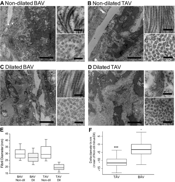
Transmission electron microscopy of collagen structure in ascending aortas. Nondilated (Non‐dil) aortas from patients with BAV (A) and TAV (B); dilated (Dil) aortas of patients with BAV (C) and TAV (D). Images represent overview, collagen fibers, and cross‐sectioned fibers. Bars are 5 μm, 200 nm, and 100 nm, respectively. Representative photomicrographs from a total of 4 patients are shown. E and F, Quantification of fibril diameters; 100 randomly selected fibrils from 4 patients were measured. ***P<0.001. BAV indicates bicuspid aortic valve; TAV, tricuspid aortic valve.