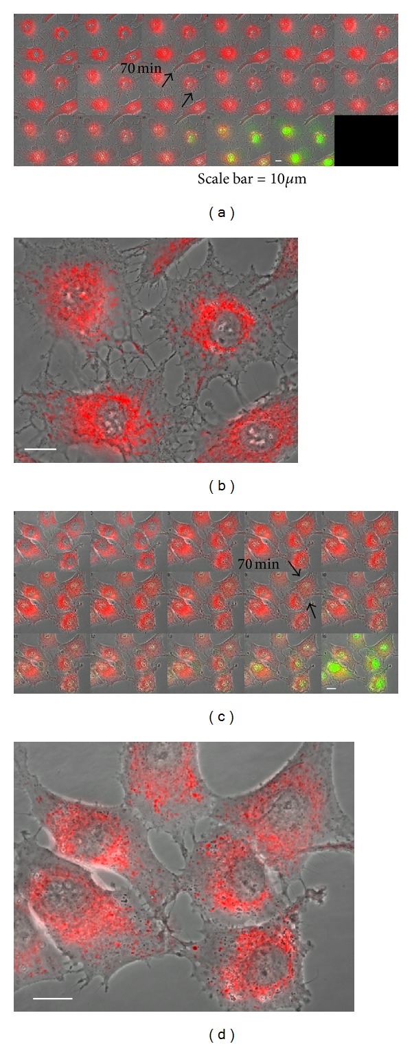
Figure 3.

RC-defective astrocytes augmented mitochondrial damage during oxidative stress-induced apoptosis. (a, b) RBA1 astrocytes and (c, d) ρ 0 RBA1 astrocytes. Time-lapse fluorescence images demonstrate dynamic changes in mitochondrial morphology using Mito-R (labeled in red and apoptotic nuclei using YO-PRO-1 (labeled in green color) during H2O2 stress. The first image of each image series in (a), (c) is the control. Dual fluorescence time-lapse images of Mito-R and YO-PRO-1 were taken simultaneously at 10 min interval for 150 min. (b), (d) Show magnification 400x of time-lapse fluorescence images at 70 mins in (a), (c), respectively. Scale bar =10 μm.
