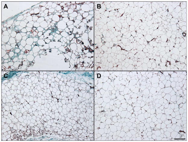
Figure 1.
Representative Histology, Masson stained. Two representative fields (100X magnification) from trichrome stained sections from each cohort are shown. Tissue from amputation patients (panels A and B) showed rare fat necrosis (panel A) amidst relatively normal adipose tissue (panel B). Tissue from control patients (panels C and D) did not show fat necrosis in the sections examined. Scale bar = 200 μm

