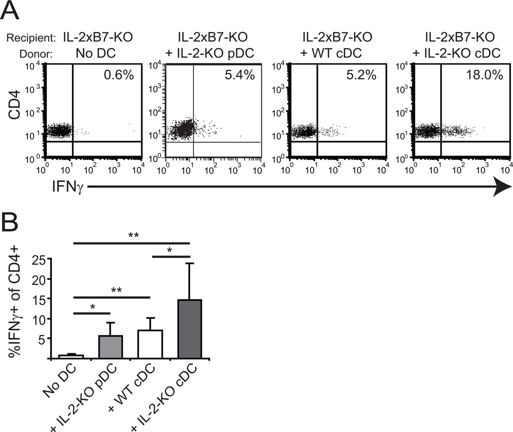
Figure 3. IL-2-KO DCs are functionally activated and induce IFNγ production by CD4+ T cells.
1×10⁶ WT or IL-2-KO cDCs or 5×10⁵ pDCs were adoptively transferred into IL-2xB7-KO recipients by tail vein injection. Injected DCs were pooled from at least three mice. Six days later, splenocytes were isolated and stimulated ex vivo with 70 ng/ml PMA and 700 ng/ml ionomycin for 6 hours with BFA added during the final 2 hours. (A) Cells were gated on CD4 and analyzed for IFNγ production by flow cytometry. (B) Bar graphs show cumulative data from three individual recipient mice and at least two experiments. *, p<0.05; **, p<0.01.

