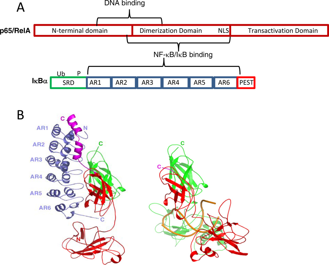
Figure 1.
(A) Schematic diagram of NF-κB(p65) one of the most abundant NF-κB family members in the cell and of IκBα, the key member of the inhibitor family. (B) LEFT: The crystal structure of IκBα (blue) bound to NF-κB (p50, green; p65, red) 11. RIGHT: The crystal structure of NF-κB (p50, green; p65, red) bound to κB site DNA (gold) 5. (Figure prepared using PyMOL 42).

