Figure 3.
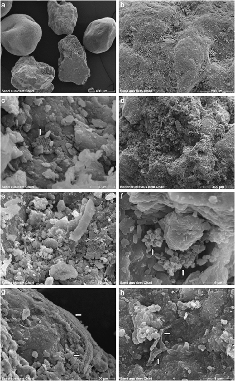
SEM micrographs of air-dried samples of surface material from Chad: Panel (a)=C2; (b)=C1; (c)=C1 magnified; (d)=C3; (e)=C4; (f)=C5; (g)=C6 and (h)=C5. Except for C3, where sand particles were held together by a thick layer of biological material, all other samples were collections of separate sand grains. A great variety of particulate matter can be seen attached to the grains: fragments of diatom shells (arrow) in (c); clumped microbial cells (arrow) in (e); collapsed microbial cells (possibly Archea – arrows in (f); mineral-encrusted microbial filaments (possibly fungal hyphae) in (g) and branched fungal hyphae (arrows) in (h). Different ratios of particulate to biogenic matter and sand grains can be seen in D, where the grains in sample C3 seem to be ‘cemented together' into a surface crust (cf samples C2 (a) and (b) (C1)).

