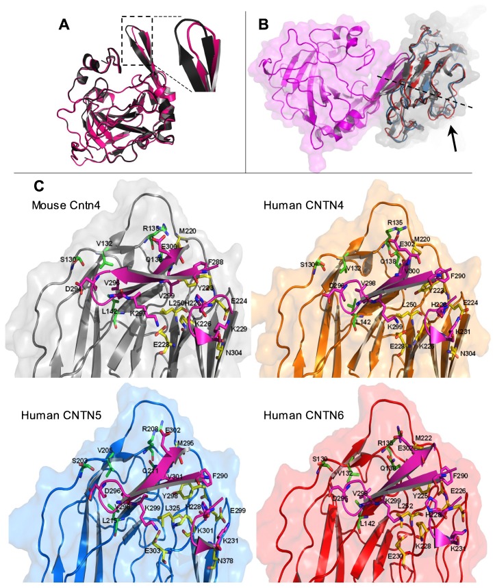
Fig. 6. Zoom magnification of the binding interfaces of the Ig-like domains of human CNTN4, -5 and -6 with the human PTPRGCA.
(A) Overlay of the carbonic anhydrase (CA) domain of mouse Ptprg with a model of the CA of human PTPRG obtained from the reference sequence of the mouse Ptprg with a specific focus on the β-hairpin (inset). Structural images were generated using PyMOL (http://www.pymol.org). (B) Alignment of the β-sheet structures within the complex between human CNTNIg1–4 and PTPRGCA symbolized with the dotted line. Arrow indicates the close vicinity of the Ω loop. (C) Comparative surface representations of the four Ig-like domains of human CNTN4, -5, and -6 with binding of PTPRG. The same orientations are represented. Residues are shown as balls and sticks. Residues from CNTNIg2, CNTNIg3 and PTPRGCA are in green, yellow, and magenta, respectively, as presented in Bouyain and Watkins for mouse Cntn4 (Bouyain and Watkins, 2010a). Cntn4 is in gray, CNTN4 in yellow, CNTN5 in blue, and CNTN6 in red. Structural images were generated using PyMOL (http://www.pymol.org).

