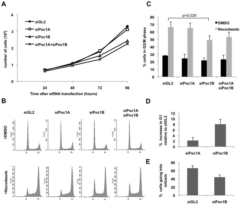
Fig. 4.
Depletion of Poc1B proteins leads to loss of proliferation. (A) Growth curves of HeLa cells transfected with siRNA oligonucleotides against luciferase as control (siGL2), Poc1A, Poc1B or Poc1A+Poc1B; data are means from two separate experiments. (B) HeLa cells treated with siRNAs for 55 hrs were treated with 0.5 µg/ml nocodazole or DMSO for 17 hours, fixed, and analysed by flow cytometry. (C) Histogram of the percentage of cells in G2/M phase; data are means ± s.d. from three separate experiments including that shown in B. The P-value indicates the significance of difference in the percentage of cells in G2/M in the nocodazole-treated siGL2 versus siPoc1B samples. (D) HeLa cells were depleted with siRNAs against GL2, Poc1A or Poc1B and analysed by flow cytometry after 72 hours. The percentage increase in G1 phase relative to siGL2 is indicated; Data are means ± s.d. from three separate experiments. (E) HeLa cells stably expressing GFP–α-tubulin were treated with siRNAs against GL2 or Poc1B for 48 hours and the percentage of cells entering mitosis in the subsequent 24 hour period was determined by time-lapse imaging. n = 113 for siGL2 and 100 for siPoc1B.

