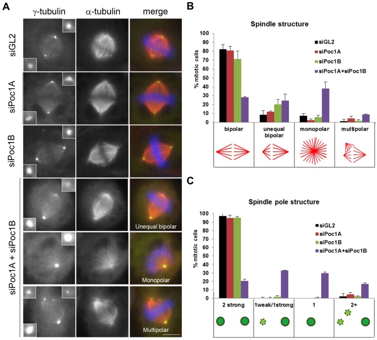
Fig. 5.
Co-depletion of Poc1A and Poc1B leads to mitotic spindle defects. (A) HeLa cells transfected with siRNAs against GL2, Poc1A and/or Poc1B were fixed after 72 hours. Cells were stained with γ-tubulin (green) and α-tubulin (red) antibodies. Merge panels include DNA staining with Hoechst 33258 (blue). Scale bar: 6 µm. (B) The percentage of cells with the spindle structures indicated. (C) The percentage of mitotic cells with γ-tubulin-positive centrosomes of the appearances indicated. Data in B and C are means ± s.d. from two separate experiments; n = 215 for siGL2, n = 218 for siPoc1A, n = 210 for siPoc1B and n = 235 for siPoc1A+siPoc1B. Error bars in B and C represent s.d.

