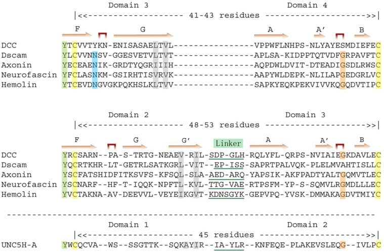
Fig. 2.
A comparative sequence alignment of peptide sequences containing D3–D4 transition (upper panel) and the D2–D3 transition (middle panel) shows the existence of a linker in D2–D3. The structure-based alignment was made by Dali pairwise comparison. Secondary structural elements are marked in accordance with the structure of DCC. A notable feature is the β turns between the F and G strands of the preceding domain and those between the A and B strands of the following domain. At the AB turn in particular there is a conserved Gly (shaded in orange), whereas N-terminal to the FG turn is a conserved Asn, which forms a complicated hydrogen bond network, exemplified by Asn178 shown in Fig. 1C. The end of the preceding domain can be defined by the alternate hydrophobic (shaded in gray) and hydrophilic residues at the C-terminus of the G strand. All of these make domain size at this area much less variable and the domain boundary easy to identify. In the D3–D4 transition, the interval between the conserved Cys (shaded in yellow) on the F strand of D3 and the Cys (shaded in yellow) on the B strand of D4 is 41–43 residues. By contrast, in the D2–D3 transition, this interval is 48–53 residues, which is indicative of a linker. Included at the very bottom of the alignment is the transition between D1 and D2 of UNC5 (bottom panel), which suggests a bend-over of D1/D2 with a five-residue linker.

