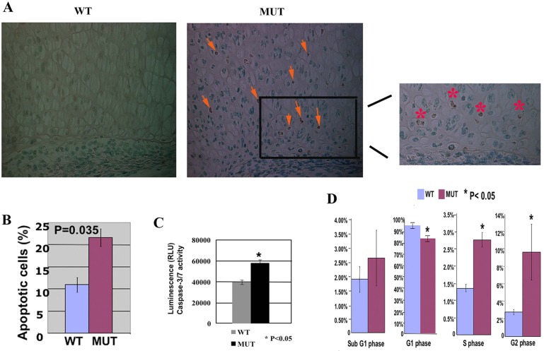
Fig. 3.
Increased apoptosis and abnormal cell cycle progression in Jab1flox/flox; Col2a1-Cre mutants. (A) TUNEL staining of apoptosis in E18.5 distal femur (40×). Significant apoptosis was detected in the mutant, whereas there was no detectable TUNEL staining in the wild-type control. Arrows point to the positive staining of apoptosis in proliferating chondrocytes. The right panel shows the enlarged image of the boxed area in the middle panel, with asterisks indicating the positive staining of apoptosis in the mutant. (B) Quantification of annexin V stained cells showed increased apoptosis in Jab1 cKO primary rib chondrocytes. (C) Increased caspase-3/7 activity in Jab1 cKO primary chondrocytes. E18.5 primary chondrocytes were treated with etoposide for 24 hours to induce apoptosis and then subjected to Caspase-Clo3/7 assay for quantification of caspase-3/7 activity as apoptosis index. (D) Quantification of flow cytometry indicates altered cell cycle progression with significant G2 phase arrest in Jab1 cKO chondrocytes. MUT, Jab1 cKO mutants. WT, wild-type littermates. n = 3 for each group.

