
Figure 2.
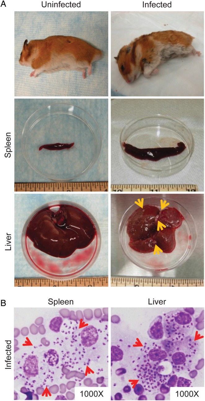
Clinicopathologic features of visceral leishmaniasis (VL) in hamsters following a vector-initiated infection with Leishmania infantum. (A) Macroscopic features of VL in a representative sick hamster (infected) compared with a healthy animal (uninfected). The infected hamster is hunched, scruffy, and thin, exhibiting dramatic weight loss (top row). The infected spleen (middle row) is enlarged, and the liver (bottom row) is fibrotic, containing multiple raised white foci (yellow arrows). The liver tissue also changes color from dark red to a brownish hue. (B) Heavy parasite burden in visceral organs of a representative infected hamster. Tissue impression smear of the spleen and liver stained with Diff-Quick shows macrophages heavily infected with L. infantum amastigotes (red arrows).