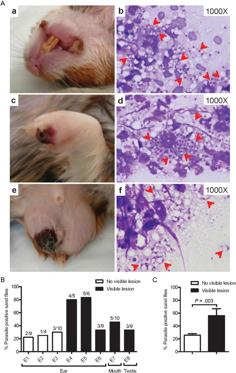
Figure 3.
Skin lesions and parasite transmissibility to sand flies in sick hamsters infected via vector-transmission of Leishmania infantum. (A) Large scabs/sores on both corners of the mouth (a), paw (c), and testis (e) from a representative infected hamster. Diff-Quick stained skin smears from mouth (b), paw (d), and testis (f) lesions showing L. infantum amastigotes (red arrows). (B) Transmissibility to sand flies from ears (original site of vector-transmission) and distal mouth and testis lesions 6–9 months following infection. Data from 8 independent exposures (E1–E8) are shown. (C) Overall success of parasite pick-up following exposure to sites with or without visible lesions. Black bars represent sites with skin lesions where sand flies were placed; white bars represent sites with no visible lesions. P < .05 was considered statistically significant.

