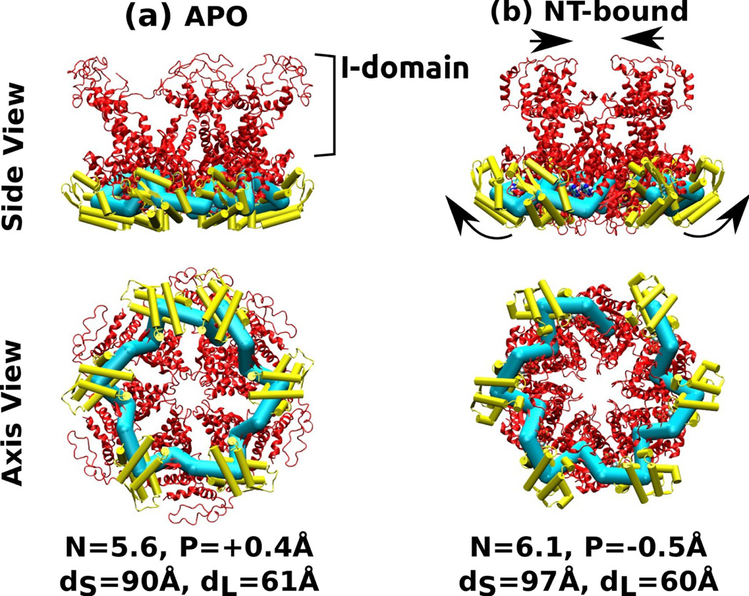
Figure 5.
Six-mer structures constructed by connecting each of the two HslU subunits in PDB 1DO2. The I-domain is attached to the large domain, and it was not used for calculating the center of mass of the large domain. (a) Nucleotide-free, (b) AMPPNP (an ATP analog) state. Arrows in (b) indicate conformational change relative to (a) that leads to widening of the small domains and narrowing of the I-domains. Symbols, molecular representations, and color codes are the same as in Fig. 4.

