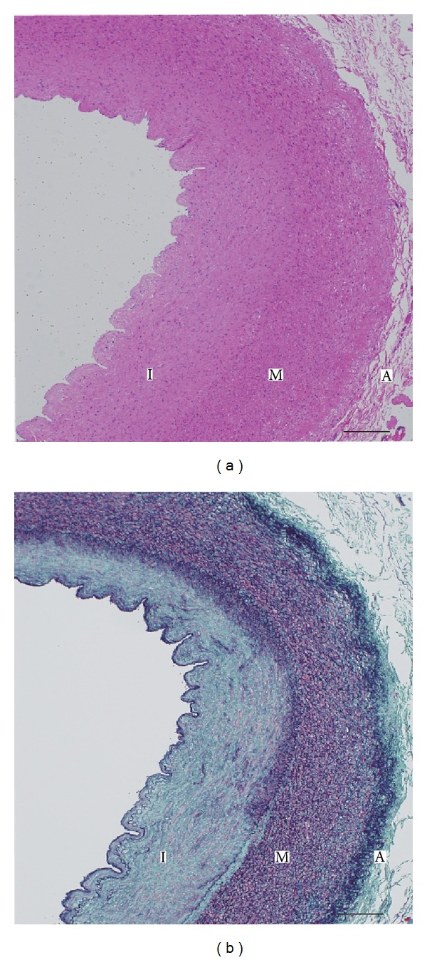
Figure 2.

Photomicrograph of thoracic aortic coarctation. Cross-sections were stained with hematoxylin-eosin (a) and Elastica-Masson (b). In panel (b), intimal proliferation (I) of collagen tissue (green) and smooth muscle (pink) is shown. Elastic fiber (purple) is decreased and substituted by collagen fiber in the outer media (M). I: tunica intima; M: tunica media; A: tunica adventitia. Scale bar: 200 μm.
