
Figure 3.
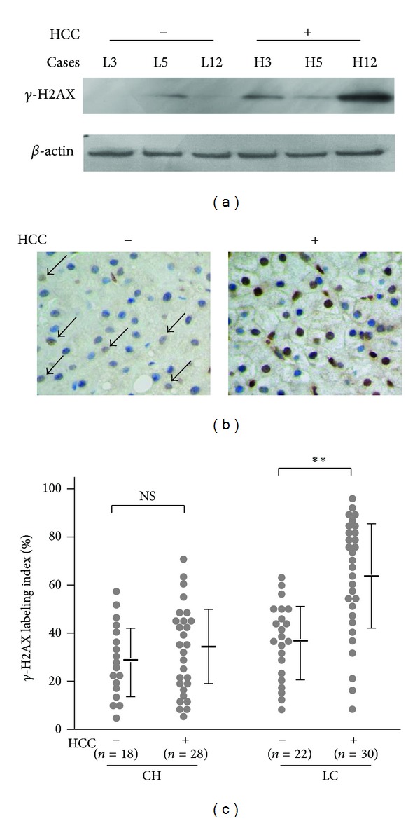
γ-H2AX is increased in the adjacent nontumorous liver tissues of HCC patients. (a) Representative data of western blotting for γ-H2AX. L3, 5, and 12: cases with liver cirrhosis without the coexistence of HCC; H3, 5, and 12: adjacent nontumorous liver tissues obtained from HCC patients. (b) Representative images of γ-H2AX immunostaining in liver tissues with and without the coexistence of HCC (original magnification ×40). (c) Dot plots showing the γ-H2AX labeling index. CH: chronic hepatitis without the coexistence of HCC (n = 18) and with HCC (n = 28); LC: liver cirrhosis without HCC (n = 22) and with HCC (n = 30). NS: not significant; **P < 0.01.