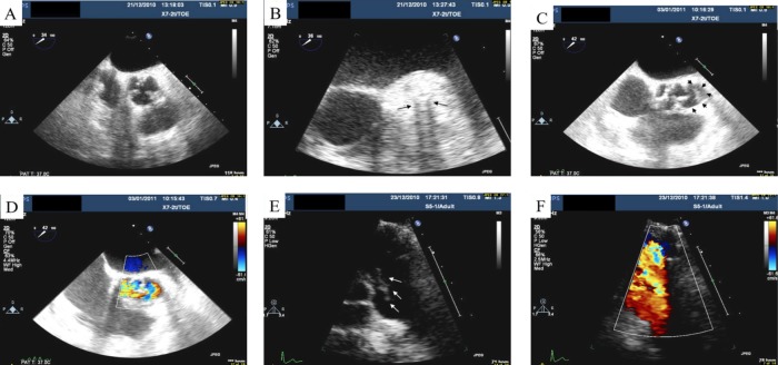
Figure 1.
(A) Transoesophageal echocardiography showing a thickening of the aortic root, most prominent laterally around the left main stem stent. (B) Zoomed image focusing on the area of thickening. Note the tramline dropout artefact relating to the stent (black arrows). (C) Black arrowheads indicate the aortic root abscess. (D) With the addition of Doppler, colour flow is seen within the abscess. (E) Transthoracic echocardiogram with white arrows indicating the vegetation on the pulmonary valve. (F) A view of the pulmonary valve with colour Doppler.

