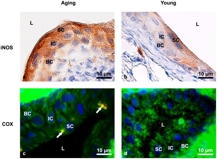
Figure 1. Immunolabeling of iNOS and COX in aging and young mice urothelium.
(a) The intense brown colour shows a strong positive immunoreaction against iNOS in the cytoplasm of superficial, intermediate and basal urothelial cells of aging mice. Nuclei are stained with haematoxylin (blue). (b) Only occasional iNOS-positive superficial cells with pale brown-coloured cytoplasm are detected in the urothelium of young mice, while intermediate and basal cells are almost completely iNOS-negative. Nuclei are stained with haematoxylin (blue). (c) Weak immunoreaction (green fluorescence) against COX in all urothelial cells of aging mice. Nuclei are stained with DAPI (blue). Arrows show lipofuscin granules. (d) Intense and extensive dotty reaction (green fluorescence) against COX in the cytoplasm of superficial urothelial cells of young mice. Nuclei are stained with DAPI (blue). Arrows show lipofuscin granules. SC-superficial cells, IC-intermediate cells, BC-basal cells, L-lumen of the urinary bladder.

