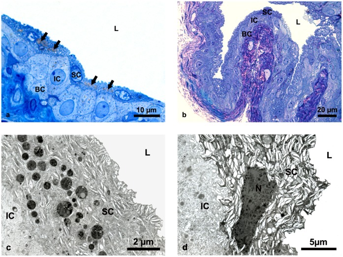
Figure 3. Structure of aging and young mice urothelium.
(a) Semi-thin section of urothelium in aging mice showing normal structure of three-layered epithelium with numerous lipofuscin granules in the cytoplasm of superficial cells (arrows). (b) Semi-thin section of urothelium in young mice showing normal structure of three-layered epithelium with no lipofuscin granules in the superficial cells. (c) Ultrathin section of aging urothelium. Ultrastructure of superficial cell fulfiled with specific fusiform vesicles and numerous osmiophilic lipofuscin granules. (d) Ultrathin section of young urothelium. Typical ultrastructural appearance of superficial cell with large amounts of fusiform vesicles in the cytoplasm but no lipofuscin granules. SC-superficial cells, IC-intermediate cells, BC-basal cells, N-nucleus, L-lumen of the urinary bladder.

