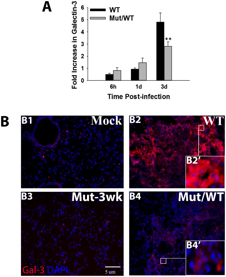
Figure 1. Upregulated expression and extracellular release of Galectin-3 in lungs during respiratory F. novicida infection.
(A) Total RNA was extracted by Trizol method from lungs harvested at the indicated times after infection with the Wild-type bacteria (WT) or from mice vaccinated with an attenuated mutant strain followed by challenge with WT bacteria (Mut/WT mice). The mRNA levels of Galectin-3 were analyzed by real-time PCR as described in Materials and Methods and are expressed as fold changes over the levels in mock control mice. Data shown are the averages of 3–4 mice per group. Statistically significant differences are denoted by asterisks (**p<0.005). (B) In-situ IF staining of frozen lung sections from mock infected and WT U112 infected or Mut/WT mice harvested at 3 d. p.i Lung harvested 3 weeks after vaccination with the mutant alone (Mut-3 wk) served as controls for Mut/WT mice. The sections were stained for galectin-3 (red) using a purified rat anti-mouse galectin-3 antibody followed by Alexa-546 conjugated chicken anti-rat antibody. Nuclei (blue) were stained with 4′6′ diamidino-2-phenylindol-dilactate (DAPI). Magnification×200. Insets depict extracellular galectin-3 in WT F. novicida infected mouse lungs (B2’) and cytosolic galectin-3 in Mut/WT (B4’) mouse lungs.

