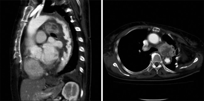
Figure 1.

Chest contrast-enhanced CT findings: an intra-aortic mass extending from the aortic arch to descending aorta and intra-aortic thrombosis was highly suspected. A mediastinal tumour, 45×40 mm, low-attenuation mass which was enhanced by contrast was also demonstrated.
