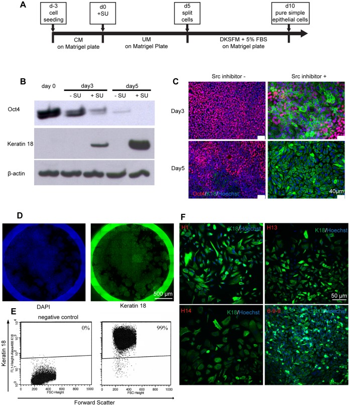
Figure 5. Modulation of β-catenin localization promotes highly efficient epithelial differentiation.
(A) Schematic of the protocol for differentiation of hPSCs to simple epithelial cells via treatment with the SFK inhibitor SU6656. (B) Western blot of protein extracts from H9 hESCs cultured for 5 days in UM without or with SU. Cytokeratin K18 protein expression was only detected in SU-treated samples. (C) Representative Oct4 (red) and K18 (green) immunocytochemistry and Hoechst (blue) staining of H9 hESCs cultured without or with SU in UM for 5 days. (D) Whole-well imaging of K18 (green) and Hoechst (blue) stained H9 cells cultured in a 12 well plate containing UM with SU for 5 days. (E) K18 flow cytometry of H9 hESCs differentiated 5 days in UM with SU. The left panel shows the negative control sample treated with the isotype control antibody. (F) Representative K18 (green) immunocytochemistry and Hoechst (blue) staining images of hESCs (H1, H13 and H14) and iPSCs (6-9-9) cultured in UM with SU for 5 days.

