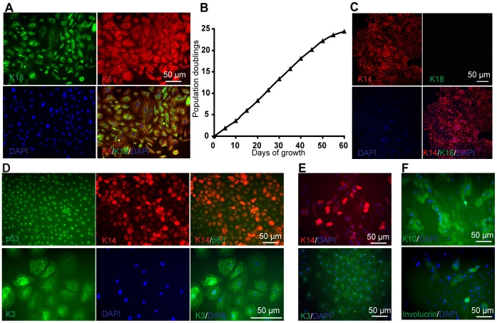
Figure 6. Expansion and terminal differentiation potential of simple epithelial cells from hPSCs.
(A) H9 cells were treated with SU for 5 days in UM. Representative K18 (green) and K8 (red) immunocytochemistry and DAPI (blue) staining are shown. (B) Subculture of H9 derived simple epithelial cells (K18+K8+) over multiple passages on 0.1% gelatin-coated 6-well plates. (C–E) The H9-derived simple epithelial (K18+/K8+) cells were treated with 1 µM RA and 10 ng/ml BMP4 for 4 days and then cultured in DKSFM for 10 days. Immunostaining of (C) K18 (green) and K14 (red), (D) p63 (green) and K14 (red), K3 (green) were performed. (E) H9-derived K18+ cells were Accutase treated to generate single cells and plated on Matrigel-coated six-well plates in DKSFM with 5% FBS at a density of 5,000 cells per well. Cells were cultured with medium changes every 7 days for 2 weeks. The resulting clones were manually picked and plated on Matrigel-coated 24-well plates at a density of one clone per well. Cells were treated with 1 µM RA for 4 days and then cultured with medium changes every 3 days for another 2 weeks. At day 30, cells were fixed and stained with K14 (red) and K3 (green) antibodies. Scale bars = 50 µm. (F) Simple epithelial cell derived K14+ cells were cultured in DKSFM supplemented with 1 mM Ca2+ for 4 days. Immunostaining of K10 (green) and Involucrin (green) were performed. Scale bars = 50 µm.

