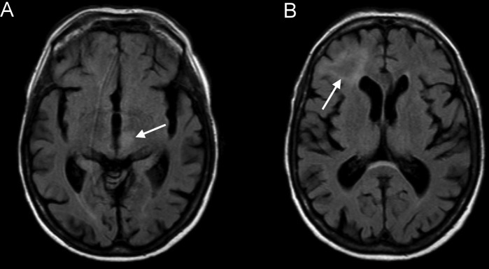
Figure 1.

Fluid-attenuated inversion recovery images approximately 3 weeks after the last rituximab administration showing hyperintense lesions in the thalamus/mesencephalon on the left (A) and in the right subcortical frontal lobe (B) without gadolinium enhancement or oedema.
