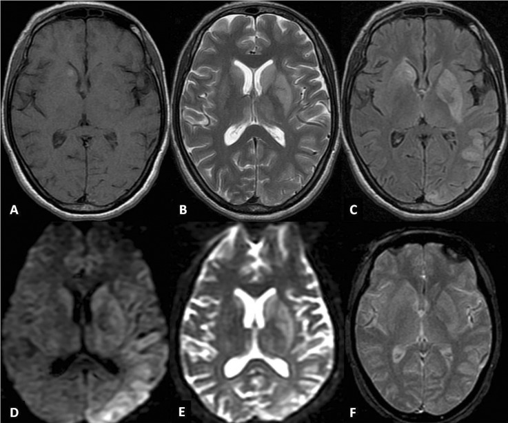
Figure 1.
MRI of the brain depicts hyperintense signal changes involving the caudate nuclei, lenticular nuclei and the thalami (left > right) on T2-weighted (B) and fluid-attenuated inversion recovery (C) images; focal hyperintensity is noted in the right caudate nuclei on T1-weighted (A) sequence. Cortical rim hyperintensity involving the posterior half of the left hemisphere is evident on T2 weighted (B), fluid-attenuated inversion recovery (C), diffusion-weighted (D) and apparent-diffusion coefficient (E) images. There is no suggestion of haemorrhage or any such transformation on gradient-recall echo (F) image.

