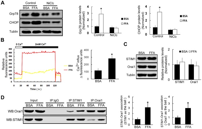
Figure 2. FFA treatments increase Ca2+ influx to induce ERS in β-TC3 cells.
(A) β-TC3 cells were incubated with FFA or BSA for 16 h, western blot was used to examine the protein expression levels of Grp78 and CHOP following the treatment with Ca2+ channel blocker NiCl2. (B) β-TC3 cells were incubated with FFA or BSA for 16 h, and then stimulated with 4 µM thapsigargin for 20 min to activate store-operated Ca2+ entry. Fluorescence densities of Ca2+ change were monitored in Fluo-8/AM-loaded β-TC3 cells after FFA or BSA treatments. (C) The protein expression levels of STIM1 and Orai1 were tested by western blot following treatments with FFA or BSA for 16 h in β-TC3 cells. (D) β-TC3 cells were incubated with FFA or BSA for 16 h, and then stimulated with 4 µM thapsigargin for 20 min. Cell lysates were immunoprecipitated with anti-STIM1 antibody followed by western blot using anti-Orai1 antibody and with anti-Orai1 antibody followed by western blot using anti-STIM1 antibody. Immunoprecipitated with anti-IgG antibody was used as the negative control. Bars represent each sample performed in triplicate, and the error bars represent the standard deviations. *P<0.05, by the Student’s t-test.

