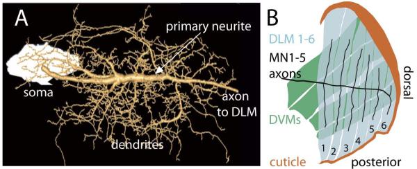
Figure 2. MN5 and the dorsal longitudinal flight muscle.

(A) Geometric MN5 dendrite reconstruction superimposed on a projection view of intracellular dye fill. The soma of MN5 is to the left (white). Soma and axon are connected by a prominent primary neurite (see white arrow), which gives rise to all dendrites. (B) Schematic drawing of inner view of the six fibers of the dorsal longitudinal flight muscle (DLM, blue) and its innervation by the flight motoneurons, MN1-5 (black). MN1-4 each innervate one of the four the ventral most DLM fibers, and MN5 innervates the two dorsal most DLM fibers. More external dorso-ventral flight muscles (DVMs) are shown in green, and the dorsal cuticle is shown in brown.
