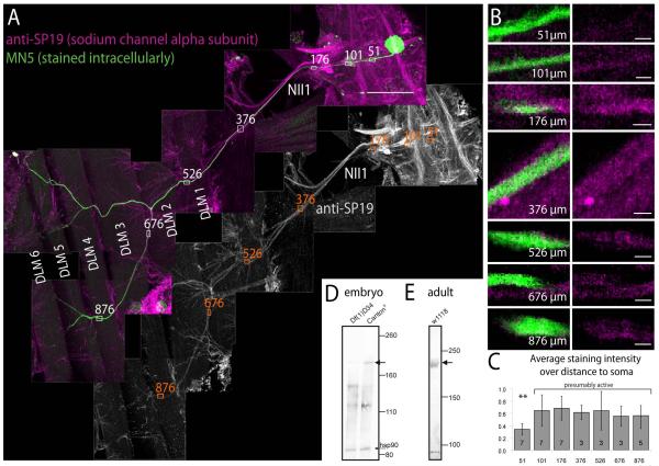
Figure 3. Localization of voltage gated sodium channels in the ventral nerve cord and in MN5.
(A) Immunohistochemistry for voltage gated sodium channels (VGSCs; magenta, upper panel; grey scale lower panel) paired with intracellular staining of MN5 (green). Several fields of view are shown as one montage that contains the pterothoracic part of the CNS, nerve NII1 to the dorsal longitudinal flight muscle (DLM), and the six fibers of the DLM (labeled DLM1-6). Within the CNS prominent axon fascicles in tracts and commissures are immunopositive for VGSCs. MN5 (green) is labeled by somatic dye injection, and its axon can be followed all the way to the two dorsal most DLM fibers (DLM5 and DLM6). Axons in nerve NII1 are immunopositive for VGSCs (see grey channel for VGSC immunostaining). Small white rectangles with numbers mark the axon of MN5 at different distances from its soma (51 μm is within the primary neurite, 101 μm is at the origin of the most distal first order dendrite, 376 μm is in nerve NII1 proximal to the DLM, 526 μm is where the NII1 reaches the DLM, and 876 μm is at just before MN5 axon branches of DLM fibers 5 and 6). (B) At each of these distances, representative single sections of MN5 and VGSC label are selectively enlarged (left column double labels, right column sodium channel immunolabel only). At 51 μm distance only faint immunopositive label for VGSCs is found in the axon of MN5 (first row). At 101 μm distance from the soma immunolabel for VGSCs in MN5 axon is stronger (second row). At all more distal locations within nerve II1A strong immunolabel for VGSCs is detected. (C) Quantification of VGSC immunostaining intensities at the distances shown in B over multiple animals (number of samples in each bar). Data were normalized to highest intensity signal in each preparation. No significant differences in VGSC immunopositive signal intensities are detected along MN5 axon between the origin of the most distal dendrite in the CNS (101 μm from MN5 soma) and the target muscle fibers DLM5-6 (ANOVA, p ≥ 0.3). By contrast more proximal parts of the MN5 primary neurite (e.g. 51 μm from the soma) show significantly lower staining intensities for VGSCs (ANOVA with Newman Keuls post hoc test, p ≤ 0.01). Scale bars: 100 μm in A, 3 μm in B. (D) Western blot from stage 16 wild type embryos (right lane) reveals a band at the size expected for Drosophila VGSC α-subunit(para, predicted size 230 kDa, see black arrow). This band is not present in stage matched para mutant embryos (Df(1)D34, left lane), see black arrow). Protein loading was similar as revealed by similarly strong bands for heat shock protein 90 (hsp90) on both lanes (see black arrow). (E) Western blot against adult brain homogenate confirms that the SP19 antibody recognized a protein of the size predicted for Drosophila VGSC α-subunit (para, predicted size 230 kDa, see black arrow).

