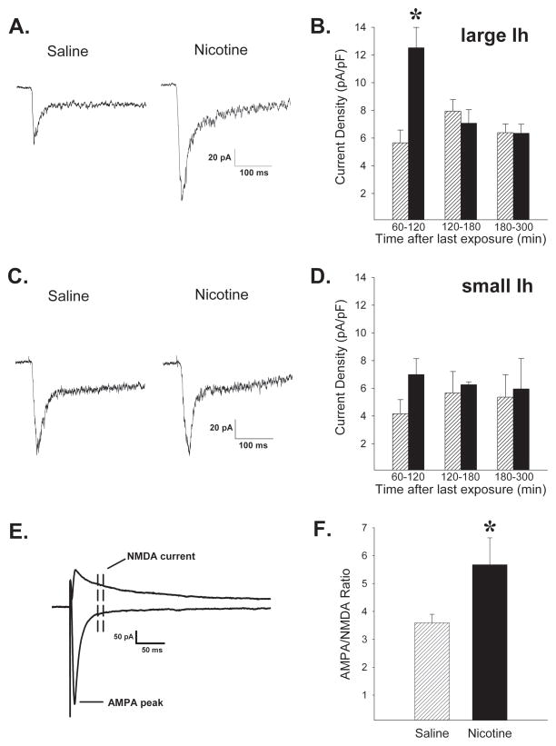
Figure 2.
Exposure to sensitizing nicotine injections (4 × 0.4mg/kg over 7 days, i.p.; ▬) leads to functional upregulation of nAChRs in VTA DA neurons and potentiation of excitatory inputs to these cells compared to saline exposed controls (
 ).Traces of nAChR current responses to 300 msec focal application of 1 mM ACh onto (A) DA (large Ih) and (C) GABA-enriched (small Ih) neurons ≤120 min after the last saline or nicotine exposure injection. B. Average peak currents normalized to cell capacitance were significantly greater in DA (large Ih) neurons of nicotine exposed rats up to 2 hours following the last nicotine exposure injection. n=38,54 cells from 10,10 rats exposed to nicotine, saline, respectively. D. No significant effects of nicotine exposure were detected in GABA-enriched neurons (small Ih). n=23,25 cells from 14,15 rats. E. Traces of evoked AMPA and NMDA EPSCs. Peak AMPA current was measured at −70 mV and the NMDA current was averaged from a window 30–40 msec after the peak of the EPSC at +40 mV. F. Mean AMPAR/NMDAR ratios were significantly greater in DA (large Ih) neurons from nicotine relative to saline exposed animals. n=13,14 cells from 8,9 rats. *, p<0.05, compared to respective saline exposure controls.

